Stem cells are a special type of cells that can
continuously proliferate in the laboratory, and can be converted into different types of
mature cells. Scientists are exploring these properties as a new form of therapy, in
which tissues and organs can be repaired or replaced by cells generated from stem
cells. But we need to know more about the biological process in which stem cells are
turned into mature cell types, in order to have better control over its effectiveness and
safety. In this project, we used a technique known as proteomics to listen to the changes
of proteins inside the cells. Since proteins are the functional molecules of cells, this
technique allows us to glimpse at the molecular makeup of stem cells during their
induced differentiation into other cell types. We have developed a new proteomic
approach that targets the newly synthesized proteins. This enables us to accurately
read which genes are turned on at different phase of stem cell differentiation. We have
applied this technique on the identification of nascent
proteins in a type of stem cells called mesenchymal stem cells. Furthermore, we have successfully
identified the nascent proteomes of Neuro2A cells during neuronal differentiation and
neurite outgrowth. In parallel, we have discovered that behaviour of mesenchymal
stem cells can be dramatically manipulated by the physical properties of the cell culture
surface. Remarkably, stem cells can be reprogrammed in vitro into differentiated
cells when exposed to a surface that mimics the corresponding tissue
microenvironment. We discovered that mesenchymal stem cells seeded on the histological section of
bovine Achilles tendon quickly adopted a highly elongated and aligned morphology,
and expressed a significantly higher level of tendon specific proteins. This suggests
that the section contains biological cues that instruct the stem cells
to commit to the tenogenic lineage. Next, we prepared polymer replicas by using
tendon sections as the mould. The resulting replica faithfully copied the physical
shape of the tendon sections. When coated with the major extracellular matrix
component of tendon, this bio-mimic culture surface promoted a similar
morphological and biochemical changes in mesenchymal stem cells that indicated a
tenocytic differentiation. This novel method opens a new horizon in stem cell biology
and tissue engineering. We invented a new material, by directly copying the
endogenous tissue environment, that can turn mesenchymal stem cells into tendon
cells. These discoveries will allow a cheaper and simpler procedure for the production of
tendon cells, and potentially other mature cell types, for cell-based therapy.
|
|
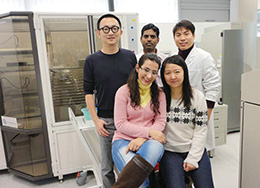
The research team led by Dr Yun-Wah Lam (back row, first from the left) applies proteomic techniques in the
understanding of fundamental cellular processes.
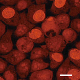
Newly synthesized proteins (labeled red) in human cells
Dr Yun-Wah LAM
Department of Biology and Chemistry
City University of Hong Kong
yunwlam@cityu.edu.hk
|